
Why Does Manual Soft-Tissue Therapy Work? The Neuroscience and Cellular Biology Behind Our Hands
Understanding mechanotransduction, sensory afferents, spinal reflexes, inflammation, and why anatomical knowledge may be the most important part of any manual technique.
For decades, manual therapists have been taught to explain the effects of soft-tissue treatment through a predominantly mechanical model.
We release fascia. We break down adhesions. We lengthen shortened muscles. We apply pressure until a Golgi tendon organ inhibits muscle activity.
These explanations are familiar, and some are useful as clinical shorthand. But when we look closely at the current research in cellular biology and neurophysiology, we find that the mechanisms behind manual therapy are considerably more complex.
And, in my opinion, far more interesting.
What we're actually doing when we apply our hands to a patient is introducing a mechanical stimulus into a living, highly innervated biological system.
That stimulus can influence sensory receptor discharge, spinal reflex excitability, nociceptive transmission, autonomic regulation, cell signalling, and potentially the extracellular matrix over time.
We're not simply changing the tissue. We're changing the information being generated by that tissue, how the nervous system processes that information, and how cells respond to mechanical loading.
This is an important distinction because it changes how we think about assessment, treatment selection, and what constitutes an effective manual intervention.
It also raises an important question.
If soft-tissue therapy works through these complicated biological mechanisms, how important is the person applying the stimulus?
I would argue that this is where clinical knowledge becomes one of the most valuable tools we have.
1. Manual Therapy Is a Form of Mechanical Neuromodulation
Every time we apply compression, stretch, shear, oscillation, or vibration to a region of the body, we mechanically deform multiple anatomical structures.
Depending on the location and direction of the force, these may include skin, superficial and deep fascia, muscle fibres, tendons, ligaments, vascular structures, and the connective tissues surrounding peripheral nerves.
All of these tissues either contain sensory receptors or exist in close mechanical relationships with structures that do.
This means a manual treatment is capable of generating substantial afferent input.
Some of that input originates from low-threshold mechanoreceptors that respond to deformation, pressure, or movement.
Other signals originate from proprioceptive systems such as muscle spindles and Golgi tendon organs.
And when tissue is injured, inflamed, or mechanically sensitized, group III and IV muscle afferents and other nociceptive free nerve endings may contribute additional information.
These signals travel through peripheral sensory neurons, their dorsal root ganglia, and into spinal cord circuitry.
From there, they may influence several interconnected systems:
Dorsal horn processing of nociceptive information.
Segmental interneuronal pathways and alpha-motoneuron excitability.
Ascending somatosensory and nociceptive pathways.
Brainstem and supraspinal pain-modulatory networks.
Autonomic and neurovascular regulation.
The key point is that these pathways are not working independently.
A change in mechanical sensory input can influence nociceptive processing and motor behaviour without requiring a permanent structural change in the tissue.
For example, a painful muscle may demonstrate increased protective activity or altered movement tolerance because of ongoing nociceptive input.
If manual treatment reduces the perceived nociceptive threat or temporarily changes reflex excitability, that patient may demonstrate improved range of motion almost immediately.
Did we lengthen the muscle?
Not necessarily.
We may have changed how the nervous system allows that muscle and joint to move.
This interpretation is supported by a 2025 living review of manual therapy mechanisms, which found evidence for peripheral, spinal, and supraspinal neurophysiological responses. Importantly, the authors also highlighted that the causal relationships between individual mechanisms and clinical outcomes remain incompletely established.
That uncertainty matters, but it doesn't make the neurology irrelevant. It means we need to become more precise about what we know and what we're still investigating.
2. Mechanotransduction: How Pressure From Our Hands Can Influence Cell Biology
One of the most fascinating developments in manual therapy research has come from the study of mechanotransduction.
Mechanotransduction is the process by which cells detect mechanical forces and convert them into biochemical signals.
Cells are not passive structures waiting to be damaged or repaired.
They continually respond to the mechanical environment around them.
When we compress, stretch, or shear tissue, those forces can be transmitted through the extracellular matrix, cell adhesion complexes, the cytoskeleton, and mechanically sensitive membrane proteins.
Integrins are particularly important because they connect extracellular-matrix structures to intracellular signalling systems.
Mechanical forces transmitted through these complexes can activate focal adhesion kinase, commonly referred to as FAK.
FAK participates in signalling networks that include extracellular signal-regulated kinases ERK1/2, which influence cellular responses to mechanical loading.
This process may affect inflammation, metabolism, extracellular-matrix regulation, and cellular adaptation.
And we have direct human evidence that massage can influence some of these pathways.
The McMaster University massage study
In 2012, Crane and colleagues published a landmark study in Science Translational Medicine examining the cellular effects of massage following exercise-induced skeletal muscle damage.
Eleven male participants underwent damaging exercise, followed by a 10-minute massage intervention applied to one quadriceps, with the other serving as a control.
Muscle biopsies were taken to examine the cellular response.
The researchers found activation of FAK and ERK1/2 signalling, increased PGC-1α-related mitochondrial biogenesis signalling, and attenuation of components of the inflammatory response, including NF-κB activation and TNF-α and IL-6 production.
These findings were important because they demonstrated that externally applied mechanical treatment could influence molecular processes within human skeletal muscle.
Interestingly, the treatment did not enhance lactate or glycogen clearance.
So the familiar explanation that massage works primarily by flushing metabolic waste is not supported by this experiment.
Instead, we have evidence that mechanically stimulating muscle can influence intracellular signalling and the response to exercise-induced damage.
There are important limitations. This was a small study of healthy participants with experimentally damaged quadriceps muscle. It does not establish that every massage technique produces the same molecular response, or that identical responses occur in fascia, tendons, ligaments, or peripheral nerves.
But it provides a compelling biological foundation.
A relatively brief mechanical intervention can influence cellular signalling long after the therapist removes their hands.
Mechanical dosage matters
Additional experimental research by Seo and colleagues in 2021 examined controlled cyclic compression in injured mouse skeletal muscle.
Appropriately dosed mechanical stimulation accelerated aspects of muscle recovery and was associated with improved clearance of neutrophils and neutrophil-derived inflammatory factors.
This is particularly interesting because neutrophils are important in the early inflammatory response to injury, yet prolonged or excessive inflammatory activity can contribute to secondary tissue damage.
The findings suggest that mechanical loading may influence the timing and character of local immune responses.
However, these were animal experiments using precisely controlled mechanical stimulation. The same biological effects have not been established for a therapist's hands in every clinical situation.
The clinical principle remains relevant.
More pressure is not automatically better.
A mechanical stimulus has an intensity, duration, frequency, direction, and rate of application. These variables help determine which biological structures are loaded and the sensory responses that follow.
We should be thinking about our hands as tools for applying a carefully selected biological stimulus, not simply generating enough force to overcome tissue resistance.
3. The Neurology of Soft-Tissue Treatment: Muscle Spindles, Golgi Tendon Organs, and Spinal Reflexes
Understanding the proprioceptive system is fundamental to understanding why manual treatment may change movement so quickly.
Two particularly important sensory systems are muscle spindles and Golgi tendon organs.
Muscle spindles: more than simple stretch detectors
Muscle spindles are specialized intrafusal receptors located within skeletal muscle, arranged in parallel with the force-generating extrafusal muscle fibres.
Their primary group Ia afferents are highly responsive to dynamic changes in muscle length, while group II and Ia afferents contribute information about sustained length.
But muscle spindle activity is not determined by mechanical stretch alone.
Its sensitivity is actively regulated by gamma-motoneuron activity, descending motor commands, task demands, and the wider sensory environment.
At the molecular level, mechanically sensitive ion channels such as PIEZO2 are fundamental to proprioceptive mechanotransduction.
Mechanical deformation can therefore be converted into changes in sensory neuron excitability and afferent discharge.
When we apply compression, shear, or vibration to muscle, we can potentially alter the mechanical conditions surrounding these receptors and the sensory information entering spinal circuitry.
However, it would be inaccurate to claim that manual therapy permanently resets muscle spindles.
That mechanism has not been directly established in humans.
Golgi tendon organs: force-dependent sensory feedback
Golgi tendon organs are located in series with muscle fibres, commonly near musculotendinous and aponeurotic regions.
Their group Ib afferents provide information related to muscle-tendon force.
Traditionally, we've been taught that stimulating these receptors creates autogenic inhibition and causes the associated muscle to relax.
The actual physiology is considerably more sophisticated.
Ib afferents project into distributed spinal interneuronal networks, where their influence depends on the movement task, mechanical context, and descending neural commands.
Their input can contribute to inhibitory or excitatory pathways.
So describing a manual treatment as simply activating a Golgi tendon organ and switching off a muscle is an oversimplification.
What we can reasonably suggest is that changing mechanical loading around a muscle-tendon unit may influence force-related sensory feedback and spinal motor control.
But the resulting response is dependent on the nervous system receiving and interpreting that information.
Evidence from H-reflex research
One of the more direct demonstrations of manual therapy influencing spinal neural function comes from research examining the Hoffmann reflex, or H-reflex.
The H-reflex is an electrically evoked response commonly used to investigate excitability in the Ia afferent–alpha-motoneuron pathway.
Research by Sullivan and colleagues, and later Goldberg and colleagues, demonstrated that massage applied to the triceps surae could substantially reduce soleus H-reflex amplitude during treatment.
This indicates a change in spinal reflex pathway excitability.
Importantly, these effects were generally transient and did not reliably persist after treatment.
The H-reflex also does not directly measure muscle spindle receptor firing. It would therefore be incorrect to interpret these findings as proof of spindle resetting.
Nevertheless, these experiments demonstrate something clinically important.
Manual mechanical stimulation can influence spinal motor circuitry within minutes, without requiring structural remodelling of the tissue.
This may contribute to why patients sometimes demonstrate immediate changes in movement resistance, perceived tightness, or available range of motion.
4. Neuroinflammation, Nociception, and the Biochemistry of Pain
Another important reason soft-tissue therapy may influence symptoms relates to the chemistry of sensitized tissues.
Pain is not simply a mechanical measurement of tissue damage.
Peripheral nociceptors respond to combinations of mechanical, thermal, and chemical stimuli.
When connective tissues or muscle become inflamed, the local environment can contain increased concentrations or activity of mediators such as bradykinin, prostaglandins, ATP, hydrogen ions, nitric-oxide-related signalling molecules, and inflammatory cytokines.
These substances can influence nociceptor excitability, sometimes lowering the threshold required to generate afferent activity.
Substance P and calcitonin gene-related peptide, or CGRP, are also important components of peripheral sensory signalling and neurogenic tissue responses.
This means that the same mechanical force may generate very different sensory responses depending on the biochemical state of the tissue.
A healthy muscle and a sensitized muscle do not necessarily respond to an identical stimulus in the same way.
From peripheral input to spinal sensitization
Nociceptive afferents originating from muscles, fascia, ligaments, and joints enter the dorsal horn of the spinal cord.
There, they interact with other sensory inputs and local interneuronal networks.
Repeated or sustained nociceptive activity may contribute to increased responsiveness within spinal nociceptive pathways.
Through convergence of sensory inputs, nociception from deep structures can also be perceived in regions beyond the exact site of origin.
This is one mechanism contributing to deep referred pain.
Low-threshold mechanoreceptive input from manual therapy enters overlapping spinal and supraspinal processing systems.
Under certain conditions, that additional input may alter nociceptive gain, motor responses, and the perceived intensity of pain.
This is not as simple as closing a single pain gate.
Descending modulatory systems involving brainstem networks, including the periaqueductal gray and rostroventromedial medulla, may also influence spinal processing.
The result is a complex interaction between peripheral sensory activity, spinal circuitry, supraspinal modulation, autonomic regulation, and the patient's current physiological state.
This provides a plausible explanation for why manual therapy can influence pain sensitivity beyond the specific tissue contacted.
It also explains why an immediate reduction in pain does not necessarily mean that the original tissue pathology has been repaired.
The neuroimmune component
The relationship between mechanical input and inflammation is another area of growing interest.
In experimental models, manual mechanical stimulation has been associated with changes in immune-cell behaviour and inflammatory mediators.
For example, a mouse study involving thoracolumbar connective-tissue inflammation identified changes in IL-4 and TGF-β1 signalling alongside differences in inflammatory markers following fascial manipulation.
These findings provide preliminary evidence for mechanobiological regulation of inflammatory processes within connective tissue.
But the direct evidence in humans is much stronger for skeletal muscle than for fascia, ligaments, tendon insertions, or other dense connective tissues.
We should not automatically translate these animal findings into claims that a particular manual technique suppresses a specific cytokine in patients.
What they do suggest is that the interface between mechanical loading, immune signalling, and neural activity deserves much greater attention in manual therapy education.
5. Fascia, Tendons, and Ligaments: Why the Tissue Still Matters
A neurological interpretation of manual therapy does not mean biomechanics are irrelevant.
The physical properties of the tissues we contact influence how applied forces are transmitted and which sensory structures may be stimulated.
Fascia contains a rich network of sensory innervation, including free nerve endings and mechanoreceptor-like structures.
Its loose connective-tissue interfaces also contain hyaluronan, which contributes to sliding between adjacent fascial layers.
Shear and compression can create reversible deformation, fluid redistribution, and changes in tissue resistance.
But there is an important difference between temporary mechanical adaptation and permanently lengthening a dense collagen structure.
Biomechanical modelling suggests that the forces required to produce meaningful plastic deformation of dense normal fascia may exceed those delivered by routine manual treatment.
So when a therapist feels a release under their hands, the experience may be genuine, but the explanation may involve changes in muscle activity, fluid redistribution, neural processing, or reversible viscoelastic properties rather than the breaking apart of collagen adhesions.
Tendons and musculotendinous junctions
The musculotendinous junction is a highly specialized interface designed to transmit force from contractile muscle into tendon.
It contains complex extracellular-matrix architecture and sensory structures involved in force feedback.
The enthesis, where tendon attaches to bone, is another mechanically specialized region with a graded transition between soft tissue and mineralized bone.
Both regions respond biologically to loading, but their adaptation is dependent on the magnitude, frequency, and duration of the applied stimulus.
Active progressive loading has substantially stronger evidence for producing clinically meaningful tendon adaptation than passive manual treatment.
This doesn't mean manual treatment around these interfaces has no value.
It means the immediate clinical benefit is more defensibly interpreted through sensory modulation, temporary mechanical changes, and improved movement tolerance, while longer-term tissue capacity should be developed through appropriately dosed loading.
This distinction is especially important in sports medicine.
Our goal should not simply be to make an athlete feel better on the treatment table.
It should be to create the conditions in which that athlete can move, load, and adapt more effectively.
6. Neurofunctional Micro Conditioning at the Tarsal Tunnel: Why Anatomy Changes Everything
Let's apply these concepts to a practical example.
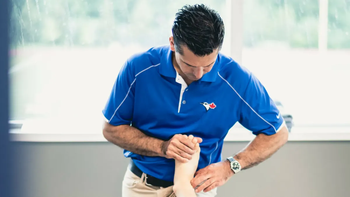
In the accompanying video, I'm performing a manual technique we call Neurofunctional Micro Conditioning in the region of the tarsal tunnel.
The tarsal tunnel is a fibro-osseous space located posteroinferior to the medial malleolus.
The flexor retinaculum forms its roof, while the underlying osseous structures create the deeper boundary.
Within this relatively confined anatomical region travel the tibialis posterior tendon, flexor digitorum longus tendon, posterior tibial artery and accompanying veins, tibial nerve, and flexor hallucis longus tendon.
The tibial nerve also gives rise to important distal branches, including the medial and lateral plantar nerves, with anatomical variation in branching patterns.
This is not simply an area of generic soft tissue.
It is an intricate anatomical interface containing tendinous, vascular, connective-tissue, and neural structures with very different mechanical and physiological properties.
What are we trying to influence?
When working around the tarsal tunnel, there are several plausible biological considerations.
Compression and shear of superficial connective tissue may provide cutaneous and fascial mechanoreceptive input.
Mechanical displacement of tendon-related tissues may change local sensory input and the loading conditions of surrounding connective tissue.
The tibial nerve and its branches are important considerations because peripheral nerves contain axons and supporting connective tissue that can be mechanically sensitive, particularly when irritated or sensitized.
Input from the region may interact with spinal sensory processing, influencing the perception of pain and potentially motor behaviour.
However, it would be incorrect to claim that a particular hand movement directly stimulates one receptor population or restores normal tibial nerve function without specific evidence.
Similarly, we cannot assume that a technique applied near the tarsal tunnel physically decompresses the nerve or produces the same biochemical changes demonstrated in studies of massaged quadriceps muscle.
Those are separate hypotheses requiring direct investigation.
The clinical question is therefore not simply whether we can apply pressure to the tarsal tunnel.
It is whether that mechanical input is appropriate for this particular patient, at this location, in the context of the findings from our assessment.
Why the therapist's anatomical knowledge is critical
Consider two therapists applying a similar-looking technique near the medial ankle.
One may simply identify an area of tenderness and apply pressure.
The other understands the relationship between the flexor retinaculum, tibial nerve, posterior tibial neurovascular bundle, flexor tendons, and their respective anatomical pathways.
They have assessed the patient's symptoms, mechanical sensitivity, neurological findings, functional limitations, and movement response.
They understand that increasing compression in a mechanically sensitive neural tunnel may not be appropriate and may aggravate symptoms.
They can modify the direction, magnitude, duration, or location of their input based on the anatomy and the patient's response.
They also understand which clinical outcome they're trying to influence and reassess whether that outcome actually changes.
From the outside, the treatment may look similar.
But the reasoning behind it is very different.
That doesn't establish that one clinician's hands produce uniquely different molecular pathways. We don't have evidence to make that claim.
It does establish why anatomical literacy, clinical judgement, stimulus selection, safety, and reassessment are central to delivering meaningful care.
The hands deliver the stimulus. The clinician's knowledge determines why, where, and how that stimulus is delivered.
7. Why the Person Performing the Treatment Matters Most
This is probably the most important clinical point I want to make.
We spend a tremendous amount of time in our professions learning techniques.
We learn where to place our hands, which direction to move, how much pressure to apply, and how long to maintain a contact.
All of those skills matter.
But the technique itself is only one part of the treatment.
The greater value lies in understanding the biological system you're attempting to influence.
What structure are you actually treating?
What is its innervation?
How does its afferent input interact with the spinal cord?
Is the presenting limitation primarily related to nociceptive sensitivity, altered motor control, reduced tissue capacity, or a combination of factors?
What mechanical stimulus makes sense given the patient's irritability and clinical presentation?
And, perhaps most importantly, how are you determining whether your treatment achieved its intended objective?
These are the questions that distinguish simply performing manual therapy from applying a clinically reasoned intervention.
I don't believe the research proves that one specific manual technique is universally superior to another.
In fact, much of the mechanistic literature suggests that different techniques may activate overlapping biological pathways.
That makes the therapist's assessment and decision-making even more important.
Because if several techniques are capable of producing a similar class of neurophysiological response, then the real clinical challenge becomes selecting an appropriate input, applying it accurately and safely, and integrating it into a treatment plan.
This is the philosophy behind how we approach neurofunctional assessment at Skills-In-Hand.
Rather than beginning with a technique and searching for somewhere to use it, we want to understand the patient's presentation, identify relevant anatomical and functional relationships, and develop a reasoned treatment strategy.
The goal is not to collect more techniques.
It is to understand why, when, and where to use the tools we already have.
As I always say, knowledge is value.
And in manual therapy, I believe anatomical and neurophysiological knowledge may be among the most valuable things we can bring to the treatment table.
8. The Future of Manual Therapy Is Mechanobiology and Neuroscience
The research surrounding manual soft-tissue treatment has evolved considerably.
We now have direct evidence that mechanically stimulating skeletal muscle can activate intracellular signalling pathways, influence inflammatory responses, and produce changes in neural excitability.
We also have evidence for short-term modulation of pain sensitivity and movement, alongside growing but less conclusive research into fascial mechanics, neuroimmune responses, and longer-term extracellular-matrix regulation.
There is still a great deal we don't know.
We cannot reliably identify which individual sensory receptor is responsible for a specific clinical outcome.
We cannot assume that a reduction in pain represents structural tissue repair.
And we do not yet have enough direct human evidence to claim that routine manual treatments regenerate tendons, permanently remodel dense fascia, or normalize peripheral nerve physiology through a specific cellular pathway.
But none of that should diminish the clinical importance of manual therapy.
It should encourage us to become more specific and more thoughtful about how we use it.
Manual therapy may create a temporary change in the biological environment. Good rehabilitation uses that opportunity to improve movement, loading, and functional capacity.
That is where the value of a knowledgeable practitioner becomes particularly clear.
A great manual therapist is not simply someone with highly skilled hands.
It's someone who understands the biology beneath those hands, can identify the relevant anatomical structures, and can apply and reassess a stimulus for a meaningful clinical reason.
That, to me, is the direction our professions should continue moving.
Key Research and References
Keter DL, et al. (2025). The mechanisms of manual therapy: A living review of systematic, narrative, and scoping reviews. PLOS ONE. Read study.
Crane JD, et al. (2012). Massage therapy attenuates inflammatory signaling after exercise-induced muscle damage. Science Translational Medicine. Read study.
Seo BR, et al. (2021). Skeletal muscle regeneration with robotic actuation-mediated clearance of neutrophils. Science Translational Medicine. Read study.
Goldberg J, et al. (1994). The effect of therapeutic massage on H-reflex amplitude in persons with a spinal cord injury. Physical Therapy. Read study.
Suarez-Rodriguez V, et al. (2022). Fascial Innervation: A Systematic Review of the Literature.Read study.
Dimitriou M. (2022). Human muscle spindles are wired to function as controllable signal-processing devices. eLife. Read study.
França MED, et al. (2020). Manipulation of the Fascial System Applied During Acute Inflammation of the Connective Tissue of the Thoracolumbar Region Affects Transforming Growth Factor-β1 and Interleukin-4 Levels.Read study.
Pethő G, Reeh PW. (2012). Sensory and signaling mechanisms of bradykinin, eicosanoids, platelet-activating factor, and nitric oxide in peripheral nociceptors. Physiological Reviews. Read study.
Davis HL, et al. (2020). Effect of sports massage on performance and recovery: a systematic review and meta-analysis.Read study.
Ahmadi M, et al. (2026). Repeated application of passive mechanical stress produces selective metabolic and extracellular matrix adaptations in human skeletal muscle but does not prevent disuse-induced atrophy.Read study.
Skills-In-Hand | Advancing clinical reasoning through anatomy, neuroscience, and neurofunctional assessment.